
The operator should use sterile gown and gloves as well. Before the procedure, the patient is placed in the Trendelenburg position. It is also important to ensure that all participants in the room are wearing a surgical mask and head cover.
Right subclavian triple lumen catheter skin#
With appropriate preparation, most subclavian venous lines can be placed without assistance however, in the operating room, a surgical scrub tech is often beneficial.īefore the initial incision or needle stick, excellent skin prep is required to prevent invocation of the catheter with skin flora. This can be completed with either betadine or chlorhexidine solution. It is important to understand the indication for the central venous catheter placement so that the correct catheter is chosen for the patient's needs.

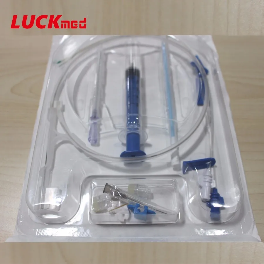
Subcutaneous ports also have different features, for example, power port capabilities to allow for rapid injection of contrast before CT scanning. More permanent tunneled catheters often have a cuff that is buried subcutaneously to prevent catheter dislodgement. Simple triple lumen catheters can have different sized lumens and can be impregnated with antimicrobial substances to prevent infection. There are also differences in catheters themselves. One will also need all supplies to ensure sterile technique throughout the procedure including skin prep, personal protective equipment, sterile draping, and dressings. General supplies for all catheter placements include a needle, guide wire, knife, dilators, and the catheter itself. Kits are available for many different needs and include triple lumen central lines, large bore catheters for dialysis, large bore single lumen "trauma" lines, permanent subcutaneous port kits and tunneled catheters. Įquipment for subclavian access depends on the type of access necessary. A potential advantage of left-sided access is the easier sweeping curve of the left innominate vein that leads to the superior vena cava located in the right mediastinum. This is importantly related to subclavian venous access because it represents another area of potential injury. The thoracic duct also terminates at the junction of the left subclavian vein and internal jugular vein. The left side pleural apex often projects more superiorly than the right leading to an increased risk of pneumothorax with left-sided access.

The subclavian vein continues beneath the clavicle heading towards the sternal notch until at the medial border of the anterior scalene muscle it joins the internal jugular vein and becomes the brachiocephalic vein, also called the innominate vein. Also important to note is the pleural apex of the lung which lies inferior to the medial aspect of the subclavian vein. Just posterior to the subclavian vein in this area is the axillary artery which becomes the subclavian artery at the lateral border of the first rib and lies in the groove of the subclavian artery. At the lateral border of the first rib, the axillary vein becomes the subclavian vein where it passes over the rib in the groove of the subclavian vein. In the normal variant of human anatomy, the subclavian vein occurs bilaterally and is a continuation of the axillary vein (a continuation of the brachial vein) from either upper extremity.


 0 kommentar(er)
